Cornea
The cornea is the outermost clear covering or window of the eye that is responsible for 60% of our eye’s focusing power. When this tissue becomes distorted, clouded, or scarred, the vision can be severely impaired.
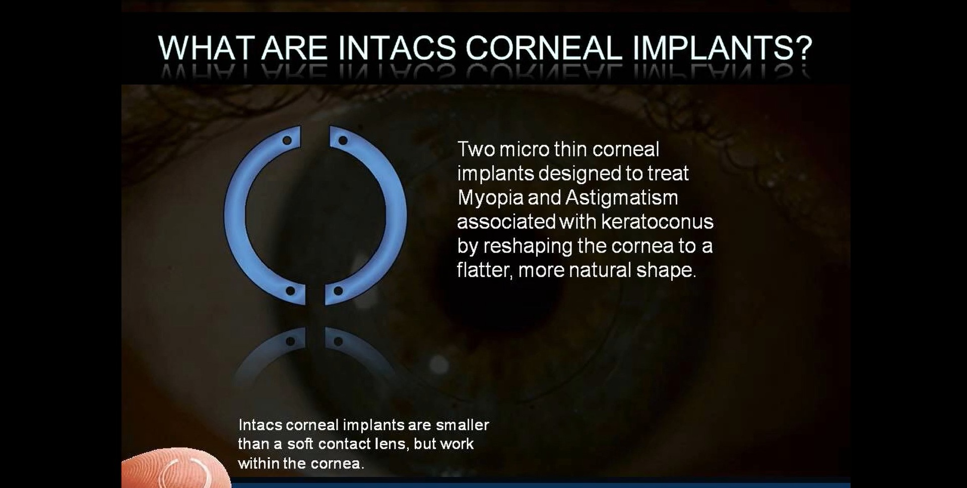
Some of the more common conditions that cause corneal problems are keratoconus (a pronounced thinning and protruding of the cornea), Fuchs’s dystrophy (a condition where there is loss of the inner layer’s cells resulting in swelling), and infections leading to scarring and opacification of the cornea tissue. Corneal problems can be visually debilitating and fairly common. A corneal transplant involves the replacement of the diseased cornea tissue with a normal donor cornea tissue from a recently deceased individual. Over 60,000 corneal transplants are performed in the USA every year.
Corneal Transplants
The donor tissue is stored in a special liquid to keep the cells viable for about a week. The donor tissue is extensively tested for clarity, cell concentration and to avoid transferring diseases such as HIV or Hepatitis. Fortunately, corneal transplant surgery can restore sight to a very good quality of vision sometimes reaching 20/20! It is not uncommon however, to require glasses or contact lenses to get the best visual result. It is performed on an outpatient basis, i.e., no hospitalization is required, and usually takes between 40 to 90 minutes varying by what type of transplant is done.
Read More
There are several types of corneal transplant surgeries depending on which part of the cornea is in need of replacement. The traditional technique involves a full thickness replacement of the defective cornea. Using a special trephine, a cookie cutter-type surgical instrument, the central portion of the diseased cornea is punched out and a “button” of cornea tissue is removed from the eye. The donor cornea is then similarly cut to fit into this space. Using approximately 16- 20 sutures that are finer than hair, the donor corneal tissue is microscopically sutured into place on the patient’s eye.
Typically, following a full thickness corneal transplant, the vision remains quite blurry for several weeks or months. Depending on the amount of astigmatism, nearsightedness or farsightedness, a person’s vision may improve faster. Patients are followed closely and the sutures are adjusted as needed to reduce astigmatism and irregularities and thus improve the vision. Eventually, most sutures will be removed but many times sutures will remain indefinitely and gradually dissolve on their own.
DSAEK
A newer technique for corneal transplantation known as Descemet’s Stripping Automated Endothelial Keratoplasty or DSAEK, began around 2005 as a minimally invasive technique to treat Fuch’s Endothelial Corneal Dystrophy. The procedure takes around 35-40 minutes and only the inner layer of the donor cornea is transplanted replacing the damaged cells of the inner layer of the patient’s cornea. This thin donor cornea tissue is gently inserted into the eye and pushed up against the inner side of the patient’s damaged cornea with an air bubble. Assured fixation of the tissue may be done with four temporary sutures to avoid early separation from the patient’s cornea and the need for further surgeries.
The DSAEK technique brings a much quicker return to good vision than the older traditional technique and is safer overall for the patient’s eye since it can be done through a tiny 3.5 mm opening. A variant of the DSAEK procedure is DMEK or Descemet’s Membrane Endothelial Keratoplasty where an even thinner layer of cells are used for the transplant. DMEK has significantly more frequent surgical and early postoperative problems requiring further surgical manipulations than does DSAEK.
Read More
Patients who undergo corneal transplant surgery usually do not encounter pain except perhaps on the first day there may be some aching of the eye. It is not uncommon to be light sensitive for about a week after the procedure and experience some watering of the eye during this time. One can often return to their usual activities a few days after the procedure.
A shield protector over the eye is commonly used during the first week primarily at night. Office visits following the procedure are typically the next day, then several days later, and then times usually double between visits. Occasionally a suture may loosen and will cause a foreign body sensation like an eyelash scratching the eye. If this happens call to be seen within 24 hours as a loosened suture can erode the delicate cornea tissue quickly and cause an ulcer. Good lubrication of the surface of the eye is required following the procedure, especially during the first few weeks. One must avoid contaminating the new tissue by washing their hands before touching their eyelids or when putting in their drops. The new vision provided by the new cornea is truly a gift of sight.

Corneal Crosslinking
Corneal crosslinking, often simply referred to as crosslinking, is a procedure that is performed to strengthen a thin or weakened cornea. The cornea may become weakened due to keratoconus, or other corneal conditions or diseases.
The procedure itself is minimally invasive, and involves utilizing liquid riboflavin, otherwise known as vitamin B2, to the cornea and then applying controlled ultraviolet light to strengthen the weakened or thin cornea.
Here at Pacific ClearVision Institute in Eugene, OR, corneal crosslinking is performed as an outpatient procedure so we can get you back to living the life you chose and stopping the progression of keratoconus so as to reduce the risk of needing a future cornea transplant.